Usted está aquí
Peruvian Journal of Neurosurgery
Endoscopy-assisted microsurgery in the resection of epidermoid tumor of the pontocerebellar angle
JERSON FLORES C., GONZALO ROJAS D., WESLEY ALABA G., RÓMULO RODRIGUEZ C.
Abstract (Spanish) ||
Full Text ||
PDF (Spanish) ||
PDF (English)
ABSTRACT
Introduction: Intracranial epidermoid tumors are rare, slow-growing, and histologically benign congenital neoplasms. The microsurgical approach with endoscopic assistance is described as a minimally invasive technique that facilitates the work of the neurosurgeon in the complete resection of an epidermoid tumor located in the cerebellopontine angle.
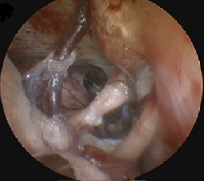
Clinical Case: 62-year-old woman, with a 3-year disease characterized by pain on the right side of the face. Symptoms increase in the following 2 years, becoming disabling and only partially improving with pregabalin and oxcarbazepine. Brain MRI showed a hypointense lesion with a cystic appearance at the level of the prepontine cistern with expansion to the right cerebellopontine angle that compressed the right trigeminal nerve (V). Retromastoid craniotomy and a right cerebellopontine angle approach were performed. Under the microscopic vision, the trigeminal nerve was identified which was pulled by the tumor, the cranial nerve complex VII-VIII, and vessels such as the superior petrous vein and the anteroinferior cerebellar artery. With the support of the endoscope, the tumor was better visualized in inaccessible areas, the prepontine cistern was accessed and total resection of the tumor was achieved. The patient evolved favorably with remission of pain on the right side.
Conclusion: The microsurgical technique assisted by endoscopy allows safe removal of the tumor, and it is immensely helpful in the resection of tumors from regions not visible under the microscope.
Keywords: Brain Neoplasms, Trigeminal Nerve, Endoscopy, Cerebellopontine Angle. (source: MeSH NLM