Usted está aquí
Peruvian Journal of Neurosurgery
Deconstructive embolization of no surgical cerebral aneurysms: a ancient solution for a complex subject
Andrés Plasencia S. MD, Alejandro Santillán MD
Abstract (Spanish) ||
Full Text ||
PDF (Spanish)
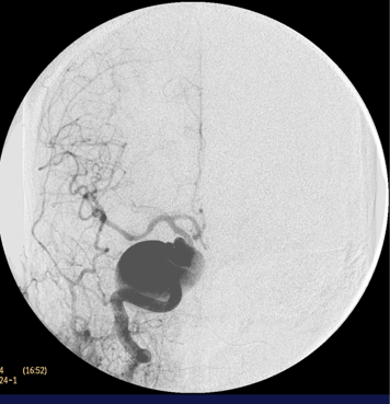
ABSTRACT
Objective.- To assess the results of endovascular deconstructive embolization of large, wide neck and fusiform aneurysms.
Methods.- We retrospectively reviewed the clinical and radiographic data of 11 cases treated between 1995 and 2009.
Results.- From the 11 aneurysms treated, 9 were saccular and 2 fusiform. The symptoms associated were mass effect in 7 of 11 (63,6%) and subarachnoid hemorrhage in 3 of 11 aneurysms (27,7%). The deconstructive approach resulted in cure for 10 patients (90,9%). There were 2 complications (18,2%), one case resulted in aphasia and hemiplegia due to deflation and migration of the balloon to the middle cerebral artery; and another case developed a delayed reversible small lateromedullary ischemia (Wallenberg syndrome). Our clinical and angiographic follow-up ranged from 6 months to 14 years (mean: 3,1 years). In all of our cured patients, the clinical improvement started few days or weeks following embolization. The remission of mass effect symptoms was achieved in all patients except in one case with previous unilateral amaurosis.
Conclusion.- Deconstructive embolization of wide neck and fusiform aneurysms still remains a useful technique in selected cases. The clinical and angiographic criteria in the balloon test occlusion seem to predict accurately the tolerance of the parent vessel for its definitive occlusion in these challenging lesions.
Key Words: Therapeutic Embolization, Saccular Aneurysm, Fusiform Aneurysm.
Spinal Metastases of Hepatocellular Carcinoma in Patient with Suspected Recurrence of Spinal Tumor
Jerson Flores C. MD, Relix Huamán H. MD, William Anicama L. MD
Abstract (Spanish) ||
Full Text ||
PDF (Spanish)
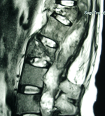
ABSTRACT
Spinal metastasis from hepatocellular carcinoma is an uncommon but serious disorder, occurring in 1.2 to 3% of patients with hepatocellular carcinoma, which is a common malignancy in developing countries and although its extrahepatic spread is common especially to lung and lymph nodes, spinal metastasis is rare and only few reports have been described in the past. The diagnosis of metastasis is made by needle biopsy in the presence of symptoms in a patient with infiltrative lesions of dorsallumbar spine and severe affectation of general condition. The treatment of spinal metastases of hepatocellular carcinoma by surgery is controversial because their appearance usually denotes the terminal state of patients many of whom are not candidates for surgery. The most widely accepted treatment option is radiation therapy, which has proved effective and safe with good symptomatic response and local control. Early diagnosis is essential for the immediate starting of radiotherapy because a positive response to radiotherapy can improve the quality of life of patients and potentially survival. We present a patient with spinal metastasis of hepatocellular carcinoma previously operated of filum terminal schwannoma.
KeyWords: Hepatocellular carcinoma; Spinal metastasis; Radiotherapy
Tension Pneumoencephalus Secondary To Frontal Sinus Osteoma: Case Report And Literature Review.
Carlos D. Fuentes V. MD, Francisco González Ll. MD, Angel Rodríguez de L. MD, Jose Hernández M. MD
Abstract (Spanish) ||
Full Text ||
PDF (Spanish)
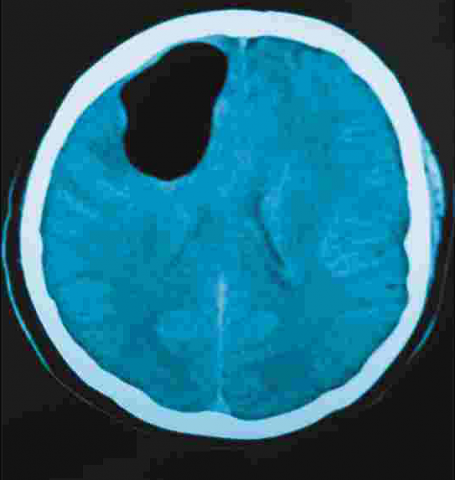
ABSTRACT
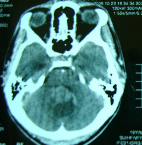
We report a 37 year-old man with tension pneumoencephalus secondary to frontal sinus osteoma. He had history of headache and left hemiparesia. CT scan revealed a large volume of air entrapped in the right frontal lobe related to an osteoma in the frontal sinus. The osteoma eroded the posterior wall of the sinus and extended into the cranial fossa. We resected the osteoma throw supraorbital craniotomy, close the dura with temporal fascia, close the bone defect with diploe and put periostium above the bone.
Surgical treatment of Pituitary Adenoma: Results in a series of 403 patients
Jerson Flores C. MD, William Lock Ch. MD, Alejandro Rosell O. MD
Abstract (Spanish) ||
Full Text ||
PDF (Spanish)
ABSTRACT
Objective: Pituitary adenomas are benign tumors that occur in approx. 10 to 15% of all intracranial tumors. Treatment can be carried by transsphenoidal resection (TSR) or transcranial resection (TCR). The aim of this study is to present the results of surgical treatment in a series of 403 patients operated at the Almenara Hospital between 2000 and 2008.
Patients and Methods: We performed a retrospective descriptive study of patients operated on pituitary adenomas from January 2000 to December 2008. It were reviewed medical records and operative reports, and gathered data on sex, age, type of adenoma, type of approach, complications, recurrence and mortality.
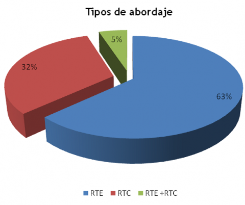
Results: 403 patients were operated of which 51% were female and 49% men. The most affected age group was 50-59 years (24.3%) followed by 40-49 years (21.2%). The most common type of adenoma was the normally functioning (60.5%). Transsphenoidal approach was used in 63%, followed by transcranial (32%) and combined (5%). TSR predominated in ACTHproducing adenomas (89%), while the TCR in prolactin-producing (70%). The most common complication was transient diabetes insipidus (50.1%). The recurrence rate was 12.9% being more frequent in the ACTH-producing tumor (21.6%) and the mortality rate was 5.2% with higher frequency in the TCR (7.0%)
Conclusions: The transsphenoidal approach the method of choice in the vast majority of pituitary tumors. The transcranial approach has specific indications and should be reserved for cases of difficult access by transsphenoidal. The adoption of new techniques, proper equipment, training and multidisciplinary working are essential to further decrease complications and mortality.
Key words: Pituitary adenoma, transsphenoidal approach, transcraneal approach
Cerebellar Mutism: Brief revision
Jose Luis Acha S. MD, Luis Contreras M. MD, Luis Huaman T. MD, Marco Chipana S. MD
Abstract (Spanish) ||
Full Text ||
PDF (Spanish)
ABSTRACT
Cerebellar mutism is a non infrequent alteration of the language in patients who underwent to a surgery of different injuries, generally tumors, affecting vermis or cerebellar hemispheres. The anatomical base of this syndrome is not well understood yet, nevertheless the injury of the dominant superior cerebellar hemisphere or the deep medial cerebellar nuclei would play an important role in the physiopathology. In this paper we make a brief revision of this problem at the time we report a case of a 18 years-old patient who had a mass in the cerebellar vermis that was operated trough a suboccipital craniotomy and telovelar approach. At the second day of postoperative period the patient developed mutism. After 16 days, the patient began to speak and improved his communication slowly.
Key Words: Cerebellar Mutism, posterior fossa tumors, posterior fossa surgery
Third Cranial Nerve Palsy as sign of Chronic Subdural Hematoma: Case Report
Jose Garcia R. MD, Elar Cari C. MD
Abstract (Spanish) ||
Full Text ||
PDF (Spanish)
ABSTRACT
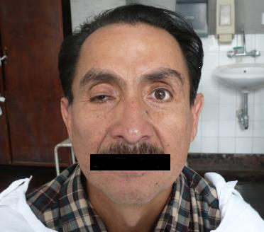
Chronic subdural hematoma (CSH) is a common pathology in elderly patients. Its etiology is usually due to rupture of bridging veins, which kept bleeding into the subdural space will generate a chronic inflammatory process which results in a collection that will grow in volume producing symptoms of intracranial hypertension or neurological focus. The third nerve palsy presenting as chronic subdural hematoma is rare, we present the case of a patient with this disease treated in our hospital.
Key Words: Hematoma subdural crónico, parálisis del tercer nervio craneal
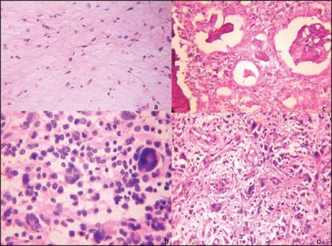
Intraventricular astroblastoma: Case report
Dante Valer G. MD, Betty Quintanilla C. MD, Jerson Flores C. MD, Alejandro Rosell O. MD, Sandro Casavilca Z. MD, Arie Perry MD
Abstract (Spanish) ||
Full Text ||
PDF (Spanish)
ABSTRACT
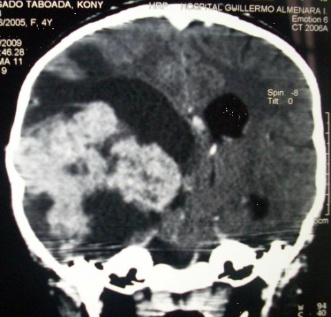
Astroblastoma is a tumor of the glial lineage of unknown origin and uncertain prognosis. Represents 0,4 - 2,8% of brain gliomas, which generally presents in youth people as an hemispheric well defined tumor, often associated with a cystic component, being extremely rare intraventricular location. The author reports the case of a patient of 4 years old girl with intraventricular astroblastoma and a review of the literature. Complete resection was demonstrated in early postoperative CT. The patient began disease since three month before the surgery with persistent headache, nausea and vomiting. She suffer cranial trauma and the indicated brain scan showed well-defined giant intraventricular tumor, at the right temporal horn with heterogeneously contrast enhancement. The patient was not treated favorably adjuvant after surgery without evidence of tumor recurrence 06 months after surgery. The histological features including nuclear pleomorphism, presence of perivascular pseudorosettes astroblastic typical and perivascular hyalinization. Have distinguished two types of astroblastoma according to histological grade; however, the prognosis in cases is not directly related to the histological type. Surgical resection remains an important prognostic factor.
Key Words: Astroblastoma, intraventricular tumor
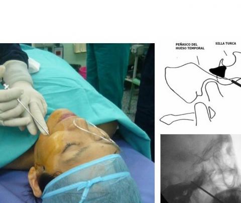
Percutaneous Rhizotomy Gasserian ganglion with ball for treatment of trigeminal neuralgia
Luis A. Huamán T. MD, Marco Gonzales-Portillo Sh. MD, Marco Chipana S. MD
Abstract (Spanish) ||
Full Text
ABSTRACT
We present four patients with trigeminal neuralgia treated by percutaneous rhizotomy of the Gasserian ganglion with balloon. The procedure was performed with fluoroscopic assistance with sedo analgesia without intubation. All patients were females. The sector most affected was V2 (50%). We obtained the three forms of balloon described (pear, hourglass, oval). In the four patients had immediate pain relief in the immediate postoperative. The procedure was ambulatory, except in one patient who was bedridden, in very poor general state of malnutrition, caused by the pain that prevented her from eating. She was discharged on the third day, eating well and walking around. One of the patients had mild dysesthesia that disappeared at the week of evolution. In all cases there was slight hypoesthesia in the hemifacie treated with progressive improvement and described as not significant for any patient.
Key words: Trigeminal neuralgia, percutaneous rhizotomy, balloon
Intraoperative rapid diagnosis by SQUASH smears in central nervous system lesions. An early institutional experience.
Bansal K K MD, Bansal Monika MD, Chuahan Neena MD, Kishore Sanjeev MD, Pathak V P MD et Al
Abstract (Spanish) ||
Full Text
ABSTRACT
Objective: Intra-operative cytological diagnosis of the CNS lesions helps the neurosurgeon to decide about the extent of resection particularly in eloquent areas. The squash or crush preparation of the available tiny tumor tissue at the start of the resection is a time saving and lead to better decision on further plan of surgery.
Material and methods - We have prospectively studied the accuracy of this technique at our institute. During the period of last two years we have operated 118 CNS mass lesions including cranial and spinal. All cases were subjected to squash diagnosis and later compared with paraffin sections.
Results - Out of total 118, cranial lesions were 105 and spinal were 13. Males outnumbered in frequency (77 cases, 65.2%) while the females comprised 41 cases (34.7%). Most (59.2%) of the patients were from 20 to 50 years of age. The cerebral hemisphere including all lobes had the largest number of cases (49 cases, 41.5%). Among them glial tumors form the major group (34 cases, 28.8%). Meningiomas were the next main group (18 cases, 15.3%) followed by Schwanommas and metastatic tumors having nine cases (7.7%) each. The cytological or squash diagnosis was possible in all except 11 cases (9.3%), in which a definite diagnosis could not be provided due to fibrous tissue, necrosis, and hemorrhage or poor preservation of cytological features.
Conclusion - Thus, our early experience concluded that Intra-operative SQUASH smear cytology is a fairly rapid and reliable method of intra operative diagnosis for a wide spectrum of central nervous system lesions.
Key words: Brain tumors, Glioma, spinal tumors, squash preparation, Neuropathology
Cervical Drezotomy to manage intractable pain in avulsion injuries of the brachial plexus
Camilo Contreras C.MD
Abstract (Spanish) ||
Full Text
ABSTRACT
Patients affected with chronic pain due to lesions by avulsion of the brachial plexus are treated with many different surgical procedures of brachial plexus and long term schemes of pain therapy remaining with high scores in VAS. DREZotomy is an important alternative to relief persistent pain related to the topography of the deafferentation. We report the results of the first cases operated in our institution by this therapy mode.
Key words: Neuropathic pain, brachial plexus avulsion, dorsal root entry
Endovascular embolization of hypervascular craniofacial tumors
Andrés Plasencia S. MD, Alejandro Santillán MD, Tomás O'Higgins MD, Trinidad Del Pino MD, Kenneth Quiroz MD
Abstract (Spanish) ||
Full Text
ABSTRACT
The autors review the most common hypervascular craniofacial tumors usually managed with endovascular embolization techniques. Even though controversial issues remain, there are reasonable data supporting the use of transcatheter devascularization techniques prior to the surgical resection of these lesions in order to reduce the intraoperative blood loss, the operative time and to facilitate their complete removal. Nevertheless, its use is not recommended as a broad treatment strategy for all craniofacial tumors. Eventually, embolization may induce and accelerate the natural involution of some hemangiomas. Randomized clinical studies must be done in order to define the efficacy and indications of these techniques.
Key words: Embolization, craniofacial tumors
Skull base approaches: Brief revision (II part)
Marco Chipana S.MD
Abstract (Spanish) ||
Full Text
ABSTRACT
The surgical approaches to reach the base of the skull represents a challenge for the neurosurgeon. The knowledge of anatomy and neurosurgical technique to use is essential before tackling the diseases that are located there. This is a continuation o a brief review of these approaches.
Key words: Skull base approaches, posterior transpetrosal approach, translabyrinthine approach, transcochlear approach, far lateral approach, transcondilar approach.
Pachymeningitis Granulomatous craneocervical in patient with suspected flat meningioma
Jerson Flores C. MD, William Lock Ch. MD , Alejandro Rosell O. MD, William Anicama L. MD
Abstract (Spanish) ||
Full Text
ABSTRACT
The hypertrophic pachymeningitis is a rare granulomatous disease of the dura, characterized by thickening of chronic granulomatous, which may affect, both intracranial and cervical or thoracic spine. Its etiology is unknown although it is associated with various concomitant disease, such as tuberculosis, AR, Wegener's granulomatosis, metabolic disorders, sarcoidosis, sinusitis, fungal infections or autoimmune disease. The differential diagnosis includes multiple meningiomas or meningiomas in plate and in the particular case of our patient, tuberculous pachymeningitis. Current treatment involves the initial rule out associated diseases such as tuberculosis because there are cases that have been referred to treatment for tuberculosis. In idiopathic cases, treatment is essentially surgical, in order to confirm diagnosis and to decompress the marrow cavity. We present a patient with hypertrophic pachymeningitis of granulomatous tuberculous etiology likely that ongoing with symptoms of multineuritis cranial and spinal cord compression, who underwent surgery in our hospital.
Key words: Idiopathic hypertrophic pachymeningitis, tuberculous pachymeningitis
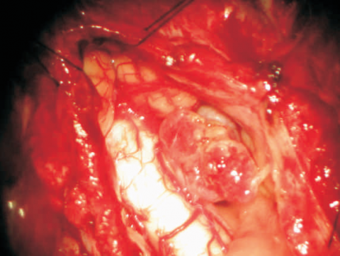
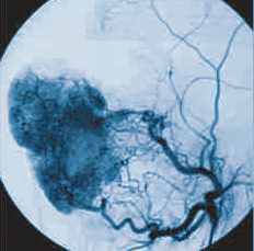
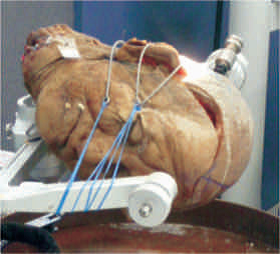
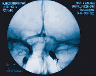
Carotid trapping as treatment for giant cervical carotid aneurysm
Jose Garcia R. MD, Ricardo Vallejos T. MD, Azucena Dávila M. MD
Abstract (Spanish) ||
Full Text
ABSTRACT
The extracranial internal carotid artery (EICA) aneurysms are very rare vascular lesions. There are series reporting incidences from 0.3 until 2% on all peripheral arterial aneurysms. Etiology has changed over the past century, from infectious cause or mycotic (tonsillar abscess and/or late manifestations of syphilis) to be less common than atherosclerotic or traumatic cause. Spontaneous dissecting cause is rarer still. Anticoagulation as first-line treatment is recommended for not broken EICA. Surgical management has been the only one method of treatment for broken EICA in the last 100 years, but was limited in those aneurysms with distal extension to the skull base. Nowadays, new technologies such as endovascular technique through the placement of stents, coils and carotid trapping are shown as alternatives for this difficult disease. We report a 44 years old man with left giant carotid cervical aneurysm, who was undertaken to carotid trapping in our hospital.
Key words: Carotid cervical aneurysm trapping
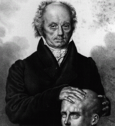
The location of the cerebral functions and the birth of Neurosurgery
Javier Torres M. MD
Abstract (Spanish) ||
Full Text
ABSTRACT
On November 25, 1884, at the Hospital for Epilepsy and Paralysis and other Diseases of the Nervous System (now Maida Vale Hospital) in London, surgeon Rickman Godlee, led by neurologists A.H. Bennet and David Ferrier, who traced in the skull the major convolutions of the Brain and the exact point where he was to trepanarse, operated from a brain tumor to the young Henderson, native of the county of Dumfries.
One hundred and twenty-five years later this seems to us of no importance and for many it will be a surprise that it wants to give the transcendence of being the birth of a new specialty: Neurosurgery.
The story is not that of an operation more to the brain, which as we know they were done from many centuries before. The transcendence is that it was the first operation of a brain tumor located only by clinical methods.