Usted está aquí
Peruvian Journal of Neurosurgery
Radiosurgery in the treatment of brain metastases
Andrés Plasencia S.MD, Aldo Berti G.MD, Mayer Saharia B.MD, Luis Pinillos A.MD, Alberto Moscol L. MD et Al
Abstract (Spanish) ||
Full Text ||
PDF (Spanish)
ABSTRACT
Radiosurgery may be chosen for brain metastases smaller than 3.5 cm. Of large diameter and with the primary tumor controlled in the following scenarios: 1) Solitary metastases and in good neurological conditions could be treated with radiosurgery only 2) Relatively radioresistant metastases such as melanoma and renal cell carcinoma, 3) Multiple metastases, complementing with total brain radiotherapy, 4) Recurrent metastases. The relative indications for radiosurgery alone, radiosurgery, more radiotherapy, radiotherapy alone and surgical resection remain controversial. Because of its proven efficacy and low morbidity, radiosurgery could be indicated for most cases of brain metastases excluding lesions larger than 3.5 cm, numerous metastases (depending on cumulative volume and whole dose to the brain) and / or with symptomatic mass effect, or causing hydrocephalus. In the latter case the surgery would be the first therapeutic consideration and when a biopsy is necessary to confirm a neoplasia.
Transcranial doppler to detect regional cerebral vasospasm in patients with SAH of aneurysmal cause.
Oscar Saldarriaga R. MD, Azucena Dávila M. MD, Fernando Palacios S. MD, Renso Trelles MD
Abstract (Spanish) ||
Full Text ||
PDF (Spanish)
ABSTRACT
Transcranial Doppler (TCD) is a useful tool in the diagnosis of both regional and diffuse vasospasm, as it is demonstrated in this case (corroborated by angiography). This makes it possible not to reduce triple therapy H when the patient still requires it, TCD is a important arsenal we have, it is not enough clinical diagnosis, since the TCD is noninvasive, fast, online, is performed in bed Patient, and the Doppler vasospasm occurs before clinical vaospasm which makes it a very useful tool. I believe that we are advancing in this area of neuromonitoring in Peru, having as final result patient with favorable evolution, if not for the measurements made.
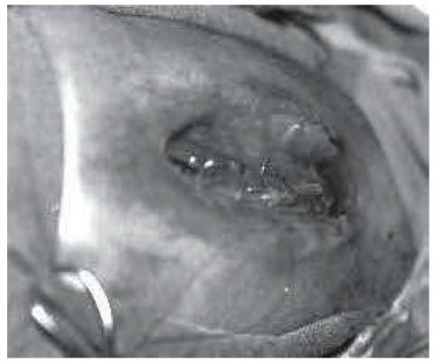
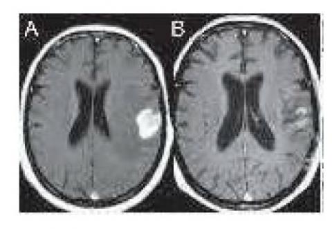
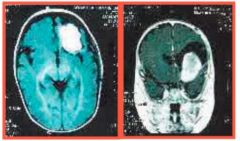
Pituitary carcinoma: Case report
Marco Chipana S. MD, Vittorio Nieri L. MD, Luis Huamán T. MD, Luis Contreras M. MD, Marco Gonzáles-Portillo S. MD
Abstract (Spanish) ||
Full Text ||
PDF (Spanish)
ABSTRACT
The pituitary carcinomas are rare and aggressive adenohypophyseal tumors, that usually present themselves like invasive macroadenomas, producing ACTH or prolactin. For clinical definition, they have metastases of a pituitary neoplasm, craniospinal or systemic. It is possible the existence of a time of latency between the diagnosis of pituitary tumor and the diagnosis of pituitary carcinoma that may vary from 5 to 12 years. The histological study is necessary for the diagnosis, and reveals functionally tumors, microscopically atypical, with high mitotic activity. The survival after documentation of metastases is low, being of only 34 % at the end of the first year. The treatmentincludes radical surgery and the use of radiotherapy and chemotherapy, nevertheless the results are still discouraging.
Key Words: Pituitary adenomas, Pituitary macroadenomas, Pituitary carcinomas.
Anaplastic ependymoma in pediatric population
Mauro Toledo MD., Ricardo Zopfi MD., Luis Gutiérrez MD., Luis Chenet MD.
Abstract (Spanish) ||
Full Text ||
PDF (Spanish)
ABSTRACT
The objective of this paper is to show the clinical presentation, neuroradiological evaluation, surgical therapy and postoperative evolution of an anaplastic ependymal brain tumor in a pediatric patient. Clinical presentation, Female patient, 8 months old, natural and from cusco, with antecedent: Craniotomy due to spontaneous intracerebral hemorrhage 4 months before admission. It enters with history of irritability, vomits, and increase of cranial volume in operative zone. Intervention and follow-up, Craniotomy was performed right temporal temporalis + Total resection of intracerebral tumor parietal temporal fronto + resection and plastid of dura mater. Neuroradiological and clinical postoperative control showed favorable evolution. Conclusion, ependymomas in the pediatric population should be considered high risk if the patient is younger than 3 years of age at the time of diagnosis, if he has preoperative metastases, features of anaplasia, or residual tumor, surgery as a therapeutic modality is the one that Provides greater disease-free survival, adjuvant therapy does not determine predictive value.
Key words: Ependymoma anaplasico, pediatric craniotomy.
History of the Discovery of Normal Pressure Hydrocephalus
Otto Campano V. MD,Nicolás Campano C. MD
Abstract (Spanish) ||
Full Text ||
PDF (Spanish)
ABSTRACT
When we read or hear Normal Pressure Hydrocephalus or chronic hydrocephalus of the adult as it is called at the moment, the Hakim Adams Syndrome comes to our mind immediately. We find it very important to expose the history of the discovery of hydrocephalus at normal pressure,
Pathology that to date has many questions and is a subject of constant research.
Radiosurgery in acoustic neurinoma: Preliminary results at the Pacific Radiosurgery Institute
Andrés Plasencia S.MD, Aldo Berti G.MD, Mayer Saharia B.MD, Luis Pinillos A.MD, Alberto Moscol L.MD et Al
Abstract (Spanish) ||
Full Text ||
PDF (Spanish)
ABSTRACT
The series of acoustic neurinoma cases treated with LINAC are limited compared to GK. Friedman et al. treated 390 patients. Actuarial control rates at 1 and 2 years were both 98% and 5% at 90%. Only 4 patients (1%) required surgery for tumor growth. Seventeen patients (4.4%) experienced facial paresis and 14 patients (3.6%) reported facial hypoesthesia. The risk of these complications is directly related to tumor volume and / or the prescribed dose at the periphery of the tumor. Since the doses were reduced to 1250 cGy in 1994, only 2 patients (0.7%) had facial paresis and another 2 (0.7%) facial numbness. The safety and efficacy of ER as an alternative to microsurgery in acoustic neurinoma is emphasized.
Cognitive Impairment of Cortical Cognitive Superior Functions in Brain Trauma
Fredy Rivera C. PS
Abstract (Spanish) ||
Full Text ||
PDF (Spanish)
ABSTRACT
The presence of neuropsychological alterations in the mental status of patients with brain traumatic injury (BTI). Are characteristic of the disease and are considered as focal symptoms or isolated deficits. In frequency in the BTI. There is a usual way of presenting cognitive impairment. The alterations of the prefrontal cortex and the irregularities of the superior order of the behavior are one of the first psychopathological manifestations of BTI. From the neuropsychological point of view, frontal lobe functions, given their high level of cortical integration and control of other cognitive functions, are difficult to evaluate directly. Some of the earliest evidence comes from the well-known case of Phineas Gage (Harlow 1848-1868), who survived the accident he suffered living until he was 38 years old with the iron rod that pierced the front of his brain and went through the Head, had no obvious neurological deficits, no sensory or motor loss, and was able to speak coherently even after the accident. As the acute effects of the injury diminished, his behavior changed completely, he showed irresponsible, disorderly. The regulation of his behavior was affected by his accident.
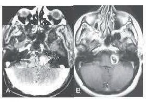
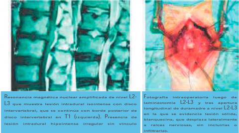
Intradural lumbar disc herniation
Marco Chipana S. MD, Eduardo Sayers C.MD, Marco Gonzáles-Portillo S. MD
Abstract (Spanish) ||
Full Text ||
PDF (Spanish)
ABSTRACT
The disc herniation is one of the most frequent pathologies in the neurosurgical practice. Localization of a portion or all of it in the intradural space is an uncommon complication that can be difficult to differentiate clinically and imagingly from both common hernias and intradural tumors. Its diagnosis is often presumptive until the moment of the surgical treatment. A clinical case of intradural lumbar disc herniation at the L2-L3 level is presented and a review of the pathogenesis, clinical features, imaging findings, treatment and prognosis of these lesions is performed.
Clinical practice guide of Brain tumours: Part III
Instituto de Enfermedades Neoplásicas, Guillermo Almenara Hospital
Abstract (Spanish) ||
Full Text ||
PDF (Spanish)
ABSTRACT
Tumors of the meninges are primary tumors that originate in meningothelial cells or non-meningothelial mesenchymal cells. The different types of existing tumors are determined by the appearance of the neoplastic cells in the microscope. 2.- Etiology: Unknown. 3.- Pathophysiology: The presence of the tumor in the intracranial level produces an imbalance of the compartments of the intracranial content, that is of the blood volume and the CSF, producing endocanean hypertension. The symptoms and signs presented are related to the topography of the lesion.4.- Epidemiology: Tumors of intracranial meninges account for 10% -15% of all primary intracranial tumors. There are no known epidemiological factors.
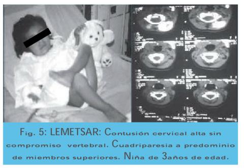
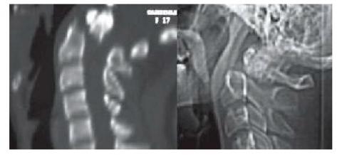
Spine cord injury in children
Germán Posadas N.MD
Abstract (Spanish) ||
Full Text ||
PDF (Spanish)
ABSTRACT
Spinal cord injury is infrequent from infancy to pre-adolescence. The incidence of traumatic lesions of the infantile spinal cord in relation to all age groups oscillates between 0.65% and 11%. Spinal injuries in relation to mild head injuries are 1 / 25-30, and severe head injuries are 1/20; However this incidence may not be as reliable because the statistics are not including minor problems or those that are not hospitalized or survive before reaching the hospital. Cirak et al. have recently studied 406 children under 14 years of age with spinal trauma, with 4% of the total number of patients admitted to the hospital.
The age of 5 to 14 years was predominant in the 36 cases studied. From the study by Cirak et al the average age was 9 years. It has been observed a higher incidence of males in a ratio of 2-3: 1 compared to the female sex; This difference is remarkable in10 years old. This is due to the greater concern, carrying out activities that demand greater risk (sports practice, driving motor vehicles, etc.) and drug consumption by older children and adolescents. On the other hand we have observed that these traumatic problems of the rachis and marrow occur mainly in the evenings and nights. Ducker and Perot report that in their cases studied at night and dawn were the predominant moments.
Direct Anterior Fixation with Cannulated Screws in Odontoid Fracture
Alfredo Fuentes-Dávila M.MD
Abstract (Spanish) ||
Full Text ||
PDF (Spanish)
ABSTRACT
The fractures of Odontoid type II according to the classification of Anderson and D'Alonzo are considered the most unstable and were always difficult to handle. External immobilization is used as a frequent method by some authors, however surgery provides better fixation. Back surgery such as inter laminar and Magerl interfacetaria are used but they block the rotational mobility of the atlas axis segment. Bohler describes the first surgeries previously, this technique was modified in recent years and is currently being performed with minimally invasive approach techniques.
In the present work we present the experience of the technique used in our series and a review of the literature with respect to this pathology and the techniques of management of such lesions.
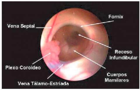
Surgical practice guide of Third Endoscopic Venriculostomy
Alberto Ramirez E.MD, Ricardo Zopli R. MD, Mauro Toledo A. MD
Abstract (Spanish) ||
Full Text ||
PDF (Spanish)
ABSTRACT
Minimally invasive surgical procedure performed through a single hole in the skull (monoportal), with the use of a neuroendoscope to allow the fenestration of the floor of the third ventricle, thus communicating the ventricular system with the subarachnoid space.
The third endoscopic ventriculostomy indications are: Obstructive or Non-Communicating Hydrocephalus (as the main indication) for: Silvio aqueduct stenosis, Tumor obstruction of cerebrospinal fluid circulation. In other types of hydrocephalus its efficacy is lower.
Clinical practice guide of Brain tumours: Part II
Instituto de Enfermedades Neoplásicas, Guillermo Almenara Hospital
Abstract (Spanish) ||
Full Text ||
PDF (Spanish)
ABSTRACT
In tumors of the posterior fossa and previous analysis of the neurological conditions upon admission (in adults, Karnofski greater than and equal to 70) and the age of the patient, a ventriculoperitoneal shunt will be planned first if there is hydrocephalus or when in Each case is justified; Then total or subtotal or partial resection of the tumor will be performed to decompress the cranial cavity, reduce endocranial hypertension in the posterior fossa, and obtain tumor material for histopathological analysis. If the tumor is located at the intraspinal level, a total or partial resection will be performed, when the patient still retains some degree of motor function in the limbs; However if there is paraplegia or quadriplegia complete grade 0/5 long installation, the operation would not be justified since it is known beforehand that motor-sensory-sphincter function will not recover. The next form of complementary treatment could be with radiotherapy for some cases with residual tumor and according to the pathology.

Epidemiology of traumatic brain injury in children
Ricardo Zopfi R.MD, Alberto Ramírez E. MD, Mauro Toledo A.MD
Abstract (Spanish) ||
Full Text ||
PDF (Spanish)
ABSTRACT
Objetive: The intention of the study was to determine the clinical and epidemiological characteristics of the patients with TCE in minors of 18 years attended in 2 hospitals of reference of Lima.
Patients and Methods: There was realized a descriptive retrospective study of 2506 patients hospitalized in 2 centers of paediatric neurosurgery: The Health National Institute of the Child and the Hospital of Paediatric Emergencies, during the year 1998 to 2002. The charts were checked and the information was grouped according to epidemiological variables as well as the characteristics of the patients with serious brain traumatic injury (TBI)
Results: It was found a predominance of the masculine sex (61%), the age group more affected was that of 1-4 years (48 %) and the fall (45 %) was one of the most frequent reasons. The symptom mas common was the vomit (61.7 %) and the sign was the cefalohematoma (23.3 %). The CT scan was done in 69 % of the patients and in 44 % of these it was found some craneal or intracraneal injury, being the most frequent diagnoses fractures of vault (30.5 %), cerebral edema (14.9 %) and epidural haematoma (12.1 %). Epidural haematoma of the convexity needed surgery in 61 % and that of posterior pit in 81 %. Acute subdural Haematoma was rare (0.08 %). The mortality in the serious BTI was very high (13 %) being the accident of traffic the most common reason (42 %) and the cerebral contusion (58.3 %) the injury most associated with mortality.
Conclusions: The BTI in the paediatric patient has special characteristic clinical and epidemiological that separate it from the adult, characteristics that must be taken in bill for the implementation of a suitable plan of attention in emergency, improvement of the reference system and of promotional preventive measurements with the purpose of decrese the morbimortalidad.
Key words: Craneal trauma in children, epidural haematoma, skull fracture
Surgical practice guide of Myelomeningocele in emergency
Luis Gutiérrez P. MD, Mauro Toledo A. MD, Ricardo Zopfi R. MD
Abstract (Spanish) ||
Full Text ||
PDF (Spanish)
ABSTRACT
Myelomeningocele is the most frequent and complex congenital malformation that is compatible with life. Our institution as a reference center at the national level for pediatric neurosurgical pathology receives with relative frequency transfers of infants with open spina bifida. According to the Office of Institutional Statistics and Informatics in Peru, during 2006, congenital malformations were the main cause of hospitalization in the neurosurgery service, with myelomeningocele being the second cause after congenital hydrocephalus.